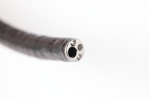
Strokes and heart attacks often strike without warning. However, a unique application of a medical camera, a scanning fiber endoscope (SFE), could one day help physicians know who is at risk for a cardiovascular event by providing a better view of potential problem areas. The scanning fiber endoscope (SFE) acquires high-quality images of atherosclerotic lesions in the carotid artery that may not be detected with conventional radiological techniques.
Developed by researchers at the University of Michigan and the University of Washington, the SFE system consists of an ultrathin, highly flexible catheter that scans blue, green, and red laser beams in a spiral pattern on the tissue surface, collecting reflectance and fluorescence images through a ring of optical fibers. The multimodal images are then reconstructed in real time with a large field of view (FOV) and projected with high spatial resolution.
The SFE generates endoscopic videos capable of discriminating early, intermediate, advanced and complicated atherosclerotic plaques. This is done by combining reflectance and laser-induced emission of intrinsic fluorescent constituents in vascular tissue.
In addition, by targeting proteolytic activity of complicated atherosclerotic plaques with a fluorescence agent activated by matrix metalloproteinases (MMPs), vulnerable regions of the weakened fibrous cap can be detected at a resolution far beyond clinically available technology.
With the complexity and accuracy required for the lens of a scanning fiber endoscope, securing the accuracy of UKA’s in-house system is the right choice. From the initial concept through final manufacturing; our team of experts is here for you.
First author Luis Savastano, MD, a Michigan Medicine resident neurosurgeon, explains that the SFE allows them to see—in very high resolution—the surface of the vessels and any lesions, such as a ruptured plaque, that could cause a stroke.
The SFE used in the study was invented and developed by co-author and University of Washington mechanical engineering research professor Eric Seibel, Ph.D. He originally designed it for early cancer detection by clearly imaging cancer cells that are currently invisible with clinical endoscopes.
According to the researchers, the superior image resolution of the technology reveals intravascular thrombi and surface thrombogenic lesions, even in cases not detected by conventional diagnostic modalities, and claim that multimodal laser-based SFE angioscopy has the potential to become a powerful platform for research, diagnosis, prognosis, and image-guided local therapy in atherosclerosis and cardiovascular disease. The study was published on February 10, 2017, in Nature Biomedical Engineering.
“The camera actually goes inside the vessels. We can see with very high resolution the surface of the vessels and any lesions, such as a ruptured plaque, that could cause a stroke,” said lead author neurosurgeon Luis Savastano, MD, of U-M. “This technology could possibly find the ‘smoking gun’ lesion in patients with strokes of unknown cause, and may even be able to show which silent, but at-risk, plaques may cause a cardiovascular event in the future.”
Universe Kogaku designs and manufactures optical lenses for scanning fiber endoscopes, security, high tech and electronic applications. We stock 1000’s of standard lens assemblies and can custom design a solution for scanners, CCTV, CCD/CMOS, medical imaging, surveillance systems, machine vision and night vision systems.