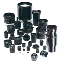
High resolution lenses for machine vision — standard and custom lens design
New 3D Imaging Software Sheds Light On Diseases
High Resolution Lenses for machine vision, instrumentation, inspection and vibration-sensitive applications. Standard and custom hi-res lens assemblies.

3D Imaging
With the development of an easy-to-use platform researchers are now able to study three-dimensional (3D) reconstruction and examine the tissues at a microscopic resolution. The new technology offers the potential to “significantly enhance the study of normal and disease processes, particularly those involving structural changes,” as reported in the American Journal of Pathology.
With 3D imaging technology, researchers have the ability to study disease manifestations, function and structure in ways that had been limited in the past because of the time, difficulty and low resolution. The most recent iteration of the 3D device was developed at the University of Leeds uses an automated virtual slide scanner as a way to generate high-resolution digital images which can then produce 3D tissue reconstructions with a cellular resolution of any tissue section.
The tissue on the slide is then digitized and scanned to the software that aligns the image and produces visualization and sends it into one virtual package. The user, and their can be multiple users, can select particular regions within the image to magnify and zoom in on; that image can then be reconfigured to produce a high-resolution image of that particular region.
In the study a mouse embryo was used to demonstrate the method could be useful for “providing anatomical and expression data and for creating a ‘virtual archive’ of 3D transgenic models.” In another test a 3D rendering of a section of the human liver showed a deposit of a colorectal carcinoma in an adjacent blood vessel that provided the researchers insight into the “tumor vasculature and its response to anti-antigenic agents.” These minute details may not have been readily visible prior to the 3D renderings and technology.
To date, the software has been found to be both robust and accurate and has the potential to increase the use of 3D histopathology as a routine research application. The advances in these technologies will lead to better and more thorough diagnosis for patients and lead to better health outcomes.